Electrical Impedance Myography
Differentiating muscle normality from abnormality.
Measures muscle composition and structure.
Building the clinical gold standard for measuring muscle quality.*
Electrical impedance myography (EIM) is a non-invasive, breakthrough technique for the assessment of muscle condition that is based on the measurement of the electrical impedance characteristics of individual muscles or groups of muscles.
A muscle’s impedance can be used to measure whether a muscle is normal (healthy) or abnormal (likely injured or diseased) by assessing the overall muscle condition and structure. These factors are characteristic of abnormality:
- Muscle fiber atrophy and disorganization
- Presence of fat and connective tissue relative to amount of muscle fibers
- Presence of inflammation
- Muscle edema
*Pending FDA clearance.

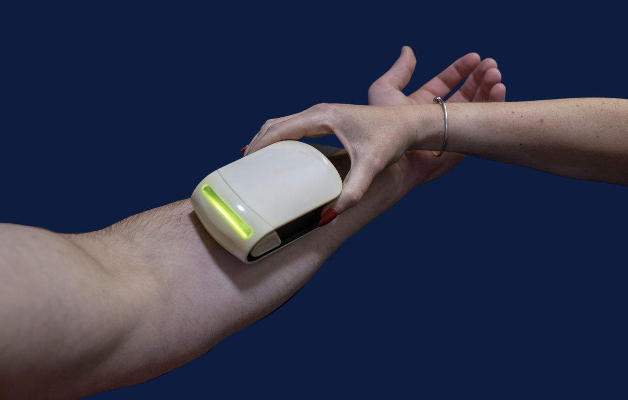
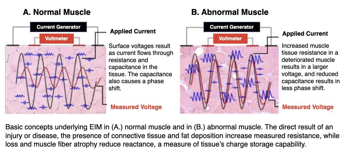
Myolex EIM assesses the electrical filtering characteristics of muscle. It is based upon the idea that a muscle’s conductivity and permittivity will become significantly altered by either injury or one of many kinds of neuromuscular or musculoskeletal disease. The integrity of individual cell membranes and alterations in the muscle’s structure and composition, including myocyte hypertrophy and atrophy, inflammation, edema, and connective tissue and fat deposition will all significantly impact the measured impedances.
Through numerous IRB-approved clinical trials, Myolex EIM has been used to help discriminate between healthy and diseased muscle, and to assess tissue degradation and progressive muscle weakness over time.
How EIM Works
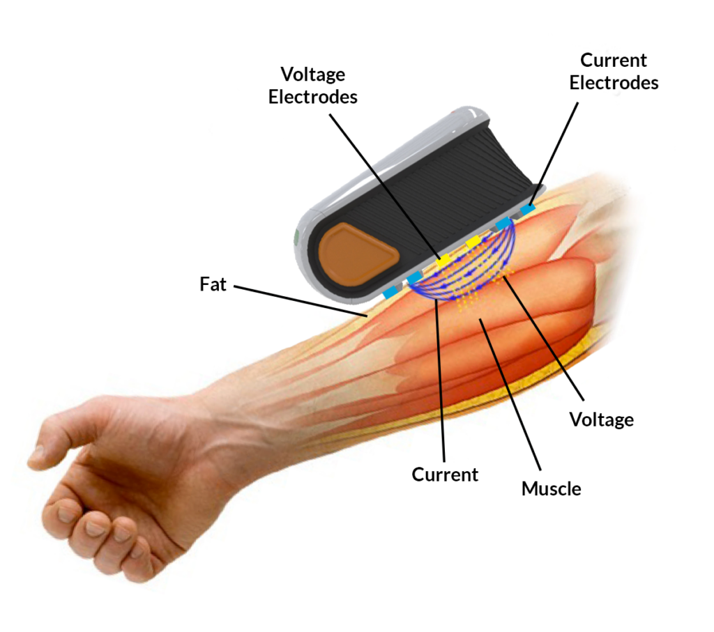
mScan measures impedance characteristics across multiple frequencies relative to the major muscle fiber direction.
Current (blue) and voltage (yellow) electrodes are placed on a specific location over a muscle. A weak electrical current is passed through the tissue across a wide range of frequencies through the current electrodes.
Alterations in the health of the muscle are reflected in the measured voltages.
App Data Display


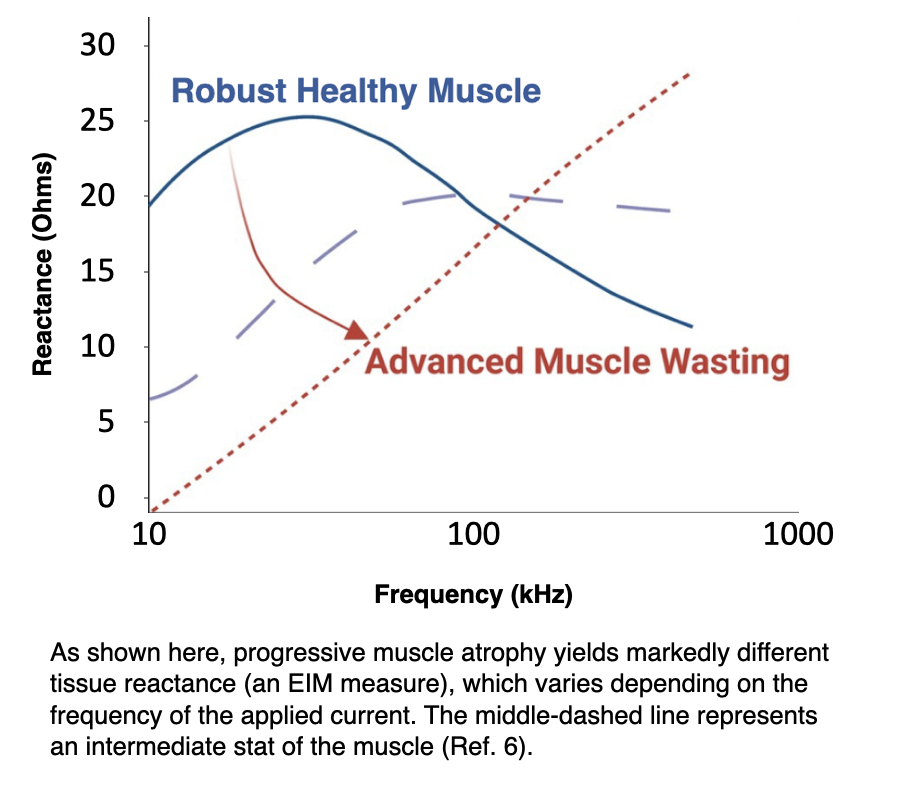
Once scanning is complete, resistance, reactance, and phase measures can be plotted as a function of frequency to demonstrate the differences in frequency dependence between healthy and degrading muscle groups.
The Five Measures of EIM
Resistance
Measures major resistance to the current through intra- and extracellular ionic fluids. Simple muscle atrophy will increase resistance of the tissue. Inflammation will cause edema in the muscle and reduce resistance.
Reactance
Highly related to myofiber membrane integrity and enables assessment of muscle fiber health. Reactance measures the additional muscle resistance due to capacitive elements such as cell membranes, tissue interfaces, and non-ionic substances. Progressive muscle atrophy yields markedly different reactance, which varies depending upon the frequency of the applied current.
Phase
For a given resistance and reactance, phase angle can then be calculated. Phase angle represents the time-shift that a sinusoid undergoes when passing through the muscle. It is less impacted by muscle size or shape.
Frequency Dependence
Different tissues (muscle, fat, bone) are uniquely sensitive to frequencies. Some tissues have great effect on one current at one frequency but not another.
Anisotropy
Skeletal muscles are unlike any other body tissue. Their conductivity and permittivity becomes significantly altered by a muscle disorder. EIM uniquely determines this anisotropy by taking muscle fiber measurements at different angles. Current flowing orthogonally across a muscle encounters more cell membranes, increasing resistance, reactance, and phase.
Patented EIM Results
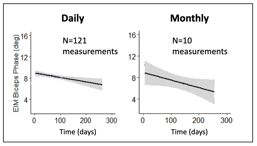
Daily, at-home measurements demonstrate greatly improved accuracy in quantifying the rate of decline in one subject’s biceps.
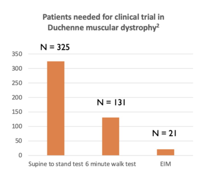
EIM detects clinical trial change with considerably fewer patients than standard measures. Also proven for ALS.

Quadriceps of 59-year-old man with ALS where disease was particularly asymmetric. Left quadriceps relatively normal. Right, markedly anomalous.
Let’s Discover Together.

